Lateral Flow Assay
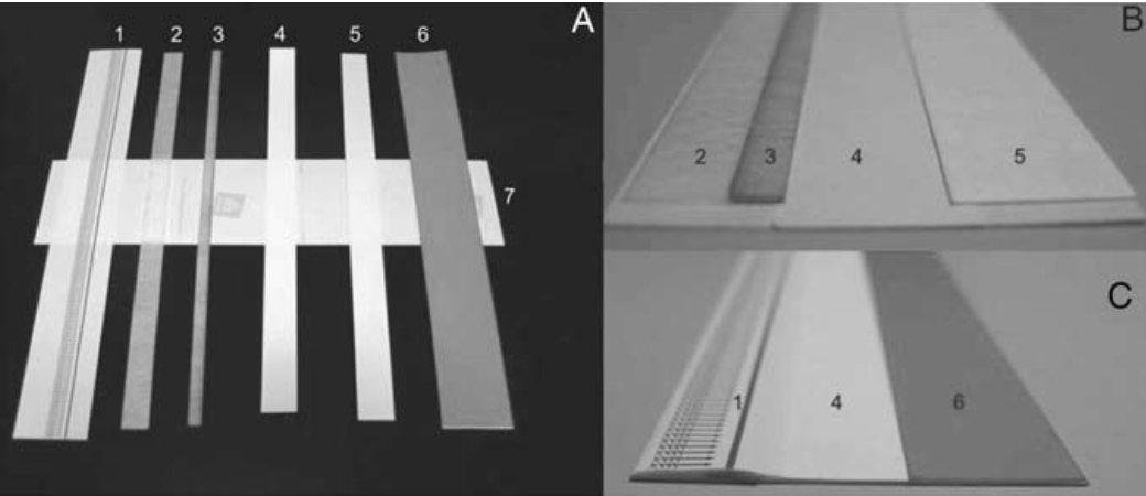
Lateral Flow Assays, also known as Lateral Flow Immunochromatographic Assays, are simple devices that detect the presence (or absence) of a target analyte within a sample, without the need for specialised and costly equipment. Numerous applications have been developed for the specific qualitative, or semi-quantitative, detection of antigens and antibodies. Typically, these tests are used for medical diagnostics either for home testing, point of care testing, or laboratory use - a widely spread, and well known, application is the home pregnancy test. The technology is based on a series of capillary beds; such as pieces of porous paper, micro-structured polymer, or sintered polymer. Each of these elements has the capacity to transport fluid (e.g., urine), spontaneously.
See below for a one-step Lateral Flow Assay protocol, which allows for the rapid identification of various analytes. The protocol describes the preparation of the assay for the detection of the Infectious Bursal Disease Virus (IBDV) and Trichinella-specific antibodies.
A. Reagents and Equipment
- Conjugate reagent: Anti-IBDV monoclonal antibody (mAb). Purify from ascitic fluids of mice carrying the specific hybridoma using a protein A column. Store at −20°C in 1 ml aliquots.
- Test line reagent: A second anti-IBDV mAb IgG recognising a different epitope of the viral protein. Purify from ascitic fluid as above and store at −20°C in 1 ml aliquots.
- Control line antibody: Goat anti-mouse IgG antibody. Isolate from sera of goats immunised with mouse IgG. Store at −20°C in 1 ml aliquots.
- Conjugate reagent: Excretory-secretory (ES) antigens. Purify from the supernatants of cultured Trichinella muscle larvae. Store at −70°C in 1 ml aliquots.
- Test line reagent: Protein A, Staphylococcus aureus (SPA) dissolved in PBS (pH 7.2) at 10 mg/ml. Store at −20°C in 1 ml aliquots.
- Control line reagent: Goat anti-ES IgG antibody. Isolate from sera of goats hyper-immunised with ES antigens. Store at −20°C in 1 ml aliquots.
Stock Solutions
- Hydrogen tetrachloroaurate (1% (w/v)): Dissolve 0.1 g of HAuCl4 in 10 ml of triple-distilled water and store in a brown bottle.
- Trisodium citrate (1% (w/v) ): Dissolve 0.5 g of Na3C6H5O7·2H2O in 50 ml of triple-distilled water.
- NaCl (1 M): Dissolve 5.8 g of NaCl in 100 ml of triple distilled water.
- K2CO3 (0.2 M): Dissolve 2.8 g of K2CO3 in 100 ml of triple-distilled water.
- Sodium borate (20 mM): Dissolve 0.76 g of Na2B4O7·10H2O in 100 ml of triple-distilled water.
- Phosphate buffered saline (PBS): Dissolve 8 g NaCl, 0.2 g KCl, 1.44 g Na2HPO4, 0.24 g KH2PO4 in 1,000 ml of triple-distilled water.
Materials and Equipment
- Nitrocellulose Membrane
- Glass Fibre Conjugate Pad Sheets (to make sample and conjugate pads).
- Filter paper (to make absorbent pads).
- Double-sided adhesive tape (to make adhesive layers on the backing plate).
- Plastic films with different colours (to make strip covers).
- Plastic boards (use for strip backing plate).
- The XYZ 3000 platform with BioJet Quanti 3000 and AirJet Quanti 3000 dispensers (BioDot Inc., Irvine, CA) is used for membrane blotting and conjugate dispensing.
- Forced Air Drying Oven (used to dry the blotted membrane and conjugate pad).
- Laminator (to assemble the master card of test strips).
- Guillotine Cutter (to cut the assembled master cards into strips).
B. Preparation and Procedure
Preparation of Colloidal Gold Suspension
- Add 1 ml of 1% (w/v) HAuCl4 solution in 100 mL of triple distilled water to a clean 500 ml Erlenmeyer flask on a stirring hot plate and bring the solution to a boil.
- To the boiling solution, quickly add 1 mL of 1% (w/v) of trisodium citrate under constant stirring.
- After the colour of the solution has changed from blue to dark red (within 3 min), continue to boil the solution for a further 2 min.
- When the solution cools to room temperature (RT), add triple-distilled water up to the original volume, stopper the flask to prevent evaporation, air circulation, and entry of contaminants and store the sample away from light.
- Scan the optical density of the red solution between 500 and 600 nm, and the absorption maximum should be at 525 nm, indicating that the gold particles have an average diameter of 40 nm.
- The colloidal gold suspension can be stored at RT in the dark for several months.
Conjugation of IgG with Colloidal Gold
for IBDV Antigen Detection | for anti-Trichinella Antibody Detection |
|
|
Membrane Blotting
for IBDV Antigen Detection |
for anti-Trichinella Antibody Detection |
|
|
Preparation of Conjugate Pads
- Cut fibreglass into 1.5× 30 cm2 strips.
- Add 1 mL of the conjugate in 2 mL of 20 mM sodium borate (pH8.0) containing 2% (w/v) BSA, 3% (w/v) sucrose, 0.6 M NaCl, 0.2% (v/v) Tween 20, and 0.1% (w/v) sodium azide.
- Put the fibreglass strips on the XYZ 3000 platform. Dispense the conjugate onto the fibreglass at 15 μl/cm using the AirJet Quanti 3000.
- Dry the conjugate pads at 50°C for 30 min using the Forced Air Drying Oven.
- Seal the strips with desiccants in a plastic bag and store at 2–8°C.
Preparation of Sample Pads
- Cut fibreglass into 1.5 × 30 cm2 strips.
- Soak the strips in PBS (pH 7.2) containing 0.1 M NaCl, 0.2% (v/v) Tween 20, and 0.1% (w/v) sodium azide.
- Dry the strips at 50°C for 30 min using the Forced Air Drying Oven.
- Seal the sample pads with desiccant in a plastic bag and store at RT.
Preparation of Absorbent Pads
- Cut filter paper in 2.5 × 30 cm2 strips.
- Seal the absorbent pads with desiccant in a plastic bag and store at RT.
Preparation of Adhesive Cards
- Put double-sided adhesive tapes on one side of the support cards.
- Cut the adhesive board into 7.5 × 30 cm2 strips to make adhesive cards.
Assembly of Master Card
- Use a laminator to or manually assemble the pre-cut materials into a lateral flow strip master card (Fig 1. A
- Stamp the blotted membrane in the middle of the adhesive backing card. Then sequentially affix the conjugate and sample pads next to the membrane with a 1–2 mm overlap at the sample end, and the absorbent pad to distal end of the membrane with 1–2 mm overlap (Fig 1. B).
- Cover the sample and conjugate pads at the sample end, and the absorbent pad at the distal end with white and blue cover films, respectively (Fig 1. C).
Cutting and Packaging
- Cut the assembled master card into 0.3 cm strips using a guillotine cutter.
- Seal the test strips with desiccants in a plastic package and store at 2–8°C.
Detection
for IBDV Antigen Detection |
for anti-Trichinella Antibody Detection |
|
|